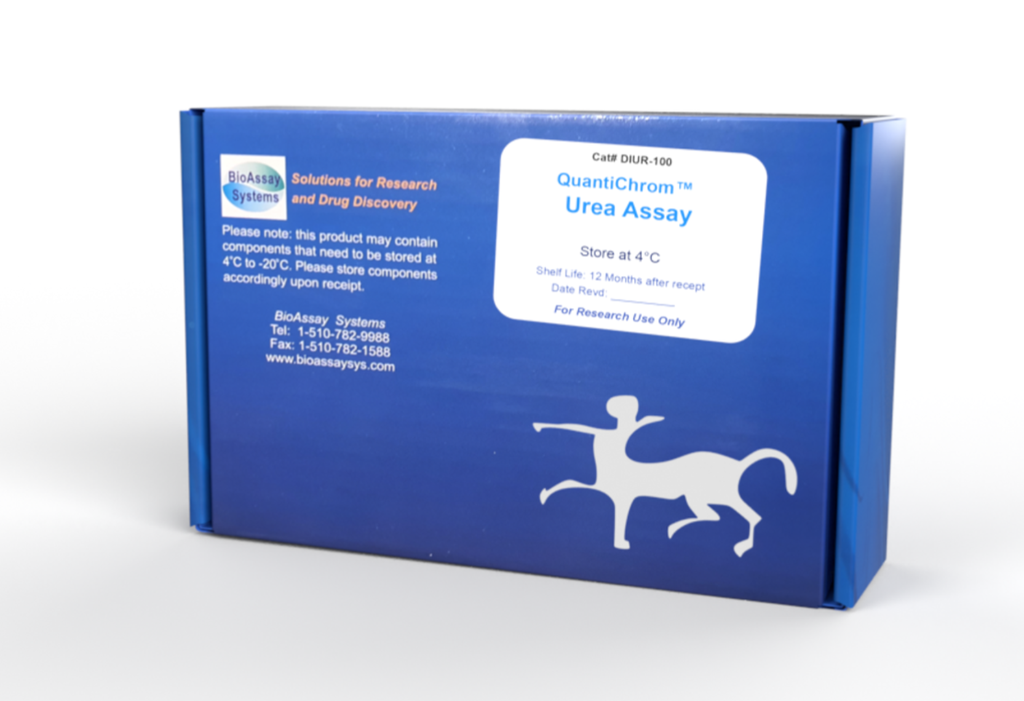
DESCRIPTION
UREA is primarily produced in the liver and secreted by the kidneys. Urea is the major end product of protein catabolism in animals. It is the primary vehicle for removal of toxic ammonia from the body. Urea determination is very useful for the medical clinician to assess kidney function of patients. In general, increased urea levels are associated with nephritis, renal ischemia, urinary tract obstruction, and certain extrarenal diseases, e.g., congestive heart failure, liver diseases and diabetes. Decreased levels indicate acute hepatic insufficiency or may result from overvigorous parenteral fluid therapy.
Simple, direct and automation-ready procedures for measuring urea concentration or blood urea nitrogen BUN in biological samples are becoming popular in Research and Drug Discovery. BioAssay Systems' urea assay kit is designed to measure urea directly in biological samples without any pretreatment. The improved Jung method utilizes a chromogenic reagent that forms a colored complex specifically with urea. The intensity of the color, measured at 520 nm, is directly proportional to the urea concentration in the sample. The optimized formulation substantially reduces interference by substances in the raw samples.
KEY FEATURES
Sensitive and accurate. Use 5 µL samples. Linear detection range 0.08 mg/dL (13 µM) to 100 mg/dL (17 mM) urea in 96-well plate assay.
Simple and high-throughput. The procedure involves addition of a single working reagent and incubation for 20 min. Can be readily automated as a high-throughput assay for thousands of samples per day.
Improved reagent stability and versatility. The optimized formulation has greatly enhanced reagent and signal stability. Cuvet or 96-well plate assay.
Low interference in biological samples. No pretreatments are needed. Assays can be directly performed on raw biological samples i.e., in the presence of lipid and protein
APPLICATIONS
Direct Assays: urea in serum, plasma, urine, milk, cell/tissue culture, bronchoalveolar lavage (BAL) etc.
Drug Discovery/Pharmacology: effects of drugs on urea metabolism.
Environment: urea determination in waste water, soil etc.
KIT CONTENTS (100 TESTS IN 96-WELL PLATES)
Reagent A: 12 mL Standard: 0.5 mL (50 mg/dL)
Reagent B: 12 mL
Storage conditions. The kit is shipped at room temperature. Store all components at 2-8°C. For long-term storage, keep standard at –20°C. Shelf life: 12 months after receipt. Precautions: reagents are for research use only. Normal precautions for laboratory reagents should be exercised while using the reagents. Please refer to Material Safety Data Sheet for detailed information.
PROCEDURES
Reagent Preparation: Equilibrate reagents to room temperature. Prepare enough working reagent for all samples and standards by combining equal volumes of Reagent A and Reagent B shortly prior to assay. Use working reagent within 20 min after mixing.
Procedure for 96-well Plate
1. Serum and plasma samples can be assayed directly (n = 1). Urine samples should be diluted 50-fold in distilled water prior to assay (n = 50). Transfer 5 µL water (blank), 5 µL standard (50 mg/dL) and 5 µL samples in duplicate into wells of a clear bottom 96-well plate. For low urea samples (< 5 mg/dL), e.g. tissue/cell extract, BAL etc, transfer 50 µL water (blank), 50 µL 5 mg urea/dL (the 50 mg/dL standard diluted 10 in water) and 50 µL samples in duplicate into separate wells. For culture medium containing phenol red, transfer 50 µL medium (blank), 50 µL 5 mg urea/dL (the 50 mg/dL standard diluted 10 in medium) and 50 µL samples in duplicate into separate wells.
2. Add 200 µL working reagent and tap lightly to mix.
3. Incubate 20 min (50 min for low urea samples) at room temperature.
4. Read optical density at 520 nm. For low urea samples, read OD at 430 nm.
Procedure for Cuvettes
Prepare samples as described for 96-well plate assay. Transfer 20 µL water, standard (50 mg/dL) and samples to appropriately labeled tubes. For low urea samples, use 5 mg/dL standard and 200 µL instead of 20 µL. Add 1000 µL working reagent and tap lightly to mix. Incubate 20 min (50 min) and read OD520nm (OD430nm).
CALCULATION
Urea concentration (mg/dL) of the sample is calculated as
ODSAMPLE, ODBLANK and ODSTANDARD are OD values of sample, blank and standard, respectively. n is the dilution factor. [STD] = 50 (or 5 for low urea samples), urea standard concentration (mg/dL).
Conversions: BUN (mg/dL) = [Urea] / 2.14. 1 mg/dL urea equals 167 µM, 0.001% or 10 ppm.
MATERIALS REQUIRED, BUT NOT PROVIDED
Pipeting devices, centrifuge tubes, Clear flat-bottom 96-well plates (e.g. Corning Costar) or cuvettes, and plate reader or spectrophotometer.
EXAMPLES
Biological samples were assayed in duplicate using the 96-well protocol. The urea concentration (mg/dL) was 12.5 ± 0.9 for Commercial 2% reduced fat milk (Kirkland), 35.7 ± 0.1 for Invitrogen fetal bovine serum, 22.1 ± 0.9 for human serum, 22.3 ± 0.2 for human plasma, 31.8 ± 1.1 for rat serum, 42.6 ± 0.1 for rat plasma and 1501 ± 52 for a fresh human urine sample, 0.21 ± 0.03 in a human BAL sample, 0.15 to 2.7 mg/dL in cell culture.
PUBLICATIONS
1. Vorland, C. J., et al (2019). Effect of ovariectomy on the progression of chronic kidney disease-mineral bone disorder (Ckd-mbd) in female Cy/+ rats. Scientific Reports, 9(1), 7936.
2. Brown, C. N., et al (2020). The effect of MEK1/2 inhibitors on cisplatininduced acute kidney injury (Aki) and cancer growth in mice. Cellular Signalling, 71, 109605.
3. Kristiansson, A., et al (2021). 177lu-psma-617 therapy in mice, with or
without the antioxidant α1-microglobulin (A1m), including kidney
damage assessment using 99mtc-mag3 imaging. Biomolecules, 11(2).
