DESCRIPTION
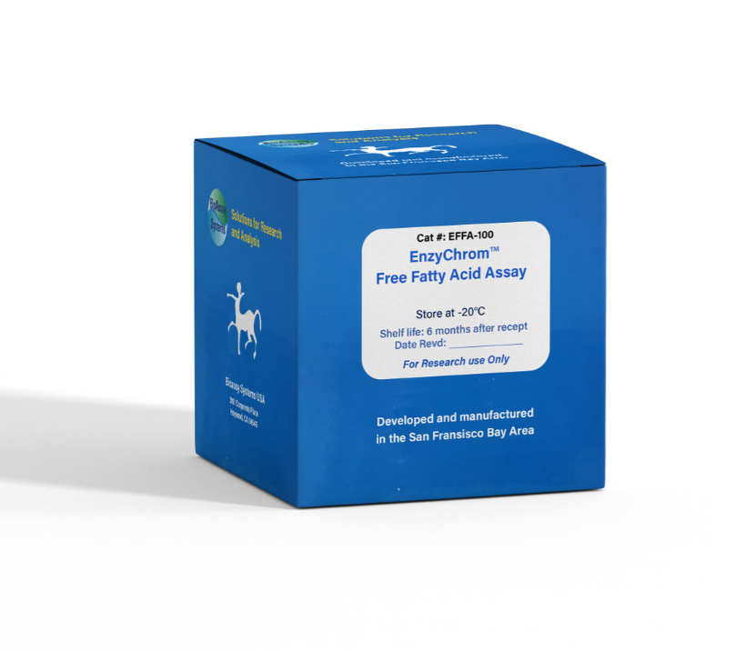
Fatty acids are aliphatic monocarboxylic acids that are ubiquitously found in animal or vegetable fat, oil and wax. Fatty Acids play important roles in cellular synthesis, energy metabolism and are implicated in diverse disorders such as diabetes mellitus, sudden infant death syndrome and Reye Syndrome. BioAssay Systems' method provides a simple, one-step and high-throughput assay for measuring free fatty acids. In this assay, free fatty acids are enzymatically converted to acylCoA and subsequently to H2O2. The resulting H2O2 reacts with a specific dye to form a pink colored product. The optical density at 570nm or fluorescence intensity (530/585 nm) is directly proportional to the free fatty acid concentration in the sample.
KEY FEATURES
Sensitive. Use 10 µL samples. Linear detection range: colorimetric assay 7 - 1000 µM, fluorimetric assay 7 - 100 µM fatty acid. Convenient. Room temperature “mix-and-read” procedure can be readily automated for high-throughput assay of thousands of samples per day.
APPLICATIONS
Assays: free fatty acids in biological samples such as serum, plasma, urine, saliva, milk, cell cultures and in food, agriculture products.
Drug Discovery/Pharmacology: effects of drugs on free fatty acid metabolism.
KIT CONTENTS
Assay Buffer: 20 mL Dye Reagent: 120 µL
Enzyme A: Dried Enzyme B: 120 µL
CoSubstrate: 120 µL Standard: 1 mL 1 mM palmitic acid
Storage conditions. The kit is shipped on ice. Store all components at -20°C. Shelf life of six months after receipt.
Precautions: reagents are for research use only. Normal precautions for laboratory reagents should be exercised while using the reagents. Please refer to Material Safety Data Sheet for detailed information.
PROCEDURES
Reagent Preparation:
Reconstitute Enzyme A by adding 120 µL dH2O to the Enzyme A tube. Make sure Enzyme A is fully dissolved by pipetting up and down and incubate at RT for 15 min. Store reconstituted Enzyme A at -20°C and use within 2 months.
Colorimetric Assay:
Liquid samples such as serum and plasma can be assayed directly. Milk and solid samples can be homogenized in 5% isopropanol and 5% Triton X-100 in water, followed by filtration through a 0.45µm PTFE syringe filter (e.g. VWR Cat# 28145-493). Note: SH-containing reagents (e.g. β–mercaptoethanol, dithiothreitol, > 5 µM), sodium azide, EDTA, and sodium dodecyl sulfate are known to interfere in this assay and should be avoided in sample preparation.
1. Equilibrate all components to room temperature. Briefly centrifuge the tubes before opening. Keep thawed tubes on ice during assay. Important: the thawed Standard solution should be clear and colorless. If it is turbid, bring it to 37°C and gently swirl the tube (do not vortex) until the solution is clear.
2. Standards: Dilute standard in Assay Buffer as follows.
Transfer 10 µL diluted standards into separate wells of a clear flatbottom 96-well plate. Samples: transfer 10 µL of each sample into separate wells of the plate.
3. Color reaction. Prepare enough Working Reagent by mixing, for each well, 90 µL Assay Buffer, 1 µL Enzyme A, 1 µL Enzyme B, 1 µL CoSubstrate and 1 µL Dye Reagent. Add 90 µL Working Reagent to each well. Tap plate to mix. Incubate 30 min at room temperature.
4. Read optical density at 570nm (550-585nm).
Fluorimetric Assay:
The fluorimetric assay procedure is similar to the colorimetric procedure except that (1) 0, 30, 60 and 100 µM Standards and (2) a black 96-well plate are used. Read fluorescence intensity at λex = 530 nm and λ em = 585 nm.
Note: if the calculated free fatty acid concentration of a sample is higher than 1000 µM in the Colorimetric Assay or 100 µM in the Fluorimetric Assay, dilute sample in Assay Buffer and repeat the assay. Multiply result by the dilution factor n.
CALCULATION
Subtract blank value (#4) from the standard values and plot the ∆OD or ∆F against standard concentrations. Determine the slope and calculate the fatty acid concentration of Sample,
RSAMPLE and RBLANK are optical density or fluorescence intensity readings of the Sample and Buffer Blank, respectively. n is the sample dilution factor.
MATERIALS REQUIRED, BUT NOT PROVIDED
Pipetting devices, centrifuge tubes, clear flat-bottom uncoated 96-well plates (e.g. VWR cat# 82050-760), optical density plate reader; black flat-bottom uncoated 96-well plates (e.g. VWR cat# 82050-676), fluorescence plate reader. For milk and solid samples, 0.45µm PTFE syringe filter and 5% isopropanol, 5% Triton X-100 solution.
PUBLICATIONS
1. Kaczmarek, A et al. (2020). The interaction between cuticle free fatty acids (Ffas) of the cockroaches Blattella germanica and Blatta orientalis and hydrolases produced by the entomopathogenic fungus Conidiobolus coronatus. PLoS ONE, 15(7).
2. Sharma, Vandana, et al (2018). Mannose Alters Gut Microbiome, Prevents Diet-Induced Obesity, and Improves Host Metabolism. Cell Reports 24.12: 3087-3098.
3. Wronska, Anna Katarzyna, et al (2018). Cuticular fatty acids of
Galleria mellonella (Lepidoptera) inhibit fungal enzymatic activities of
pathogenic Conidiobolus coronatus. PloS one 13.3: e0192715.
