| Category | Monoclonal Antibody |
| Clone Nr. | (#9G33) |
| Isotype | IgG2 |
| Species Reactivity | Mouse |
| Formulation | lyophilized |
| Buffer | PBS |
| Reconstitution | Centrifuge the vial prior to opening. Reconstitute the antibody with 200 µl sterile PBS and the final concentration is 500 µg/ml. |
| Stability and Storage | Lyophilized samples are stable for 2 years from date of receipt when stored at -20°C. Reconstituted antibody can be aliquoted and stored frozen at < -20°C for at least six months without detectable loss of activity. |
| Preparation | This antibody was produced from a hybridoma (mouse myeloma fused with spleen cells from a rat immunized with pur ified mouse PlGF2 recombinant protein. The IgG fraction of the culture supernatant was pur ified by Protein G affi nity chromatogr |
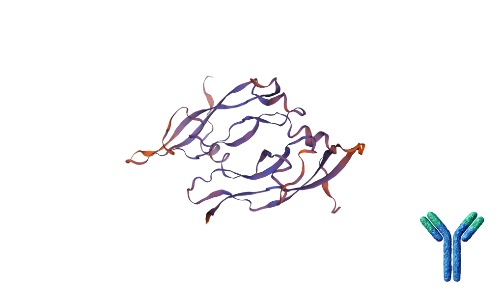
| Antigen | recombinant mouse PlGF |
| Application | WB, N |
| Synonyms | Pgf; PlGF; Plgf; AI854365; placental growth factor |
| Description | Placenta growth factor (PlGF) is a member of the PDGF/VEGF family of growth factors that share a conserved pattern of eight cysteines. Alternate splicing results in at least three human mature PlGF forms containing 131 (PlGF-1), 152 (PlGF-2), and 203 (PlGF-3) amino acids (aa) respectively. Only PlGF-2 contains a highly basic heparin-binding 21 aa insert at the C-terminus. In the mouse, only one P lGF that is the equivalent of human PlGF-2 has been identified. Mouse PlGF shares 60%, 92%, 62% and 59% aa identity with the appropriate isoform of human, rat, canine and equine PlGF. PlGF is mainly found as variably glycosylated, secreted, 55 - 60 kDa disulfide linked homodimers. Mammalian cells expressing PlGF include villous trophoblasts, decidual cells, erythroblasts, keratinocytes and some endothelial cells. Circulating PlGF increases during human pregnancy, reaching a peak in mid-gestation; this increase is attenuated in preeclampsia. However, deletion of PlGF in the mouse does not affect development or reproduction. Postnatally, mice lacking PlGF show impaired angiogenesis in response to ischemia. PlGF binds and signals through VEGF R1/Flt-1, but not VEGF R2/Flk-1/KDR, while VEGF binds both but signals only through the angiogenic receptor, VEGF R2. PlGF and VEGF therefore compete for binding to VEGF R1, allowing high PlGF to discourage VEGF/VEGF R1 binding and promote VEGF/VEGF R2-mediated angiogenesis. However, PlGF (especially human PlGF-1) and some forms of VEGF can form dimers that decrease the angiogenic effect of VEGF on VEGF R2. PlGF-2, like VEGF164/165, shows heparin-dependent binding of neuropilin (Npn)-1 and Npn-2 and can inhibit nerve growth cone collapse. PlGF induces monocyte activation, migration, and production of inflammatory cytokines and VEGF. These activities facilitate wound and bone fracture healing, but also contribute to inflammation in active sickle cell disease and atherosclerosis. Circulating PlGF often correlates with tumor stage and aggressiveness, and therapeutic PlGF antibodies are being investigated to inhibit tumor growth and angiogenesis. The functional antibody will block the binding of PlGF to Flt-1/VEGFR-1 |
| Uniprot ID | P49764 |
| Protein RefSeq | NP_032853 |
| mRNA RefSeq | NM_008827 |
